Twice a day the isotopes arrive at the Nuclear Medicine Department at Richmond Hospital.
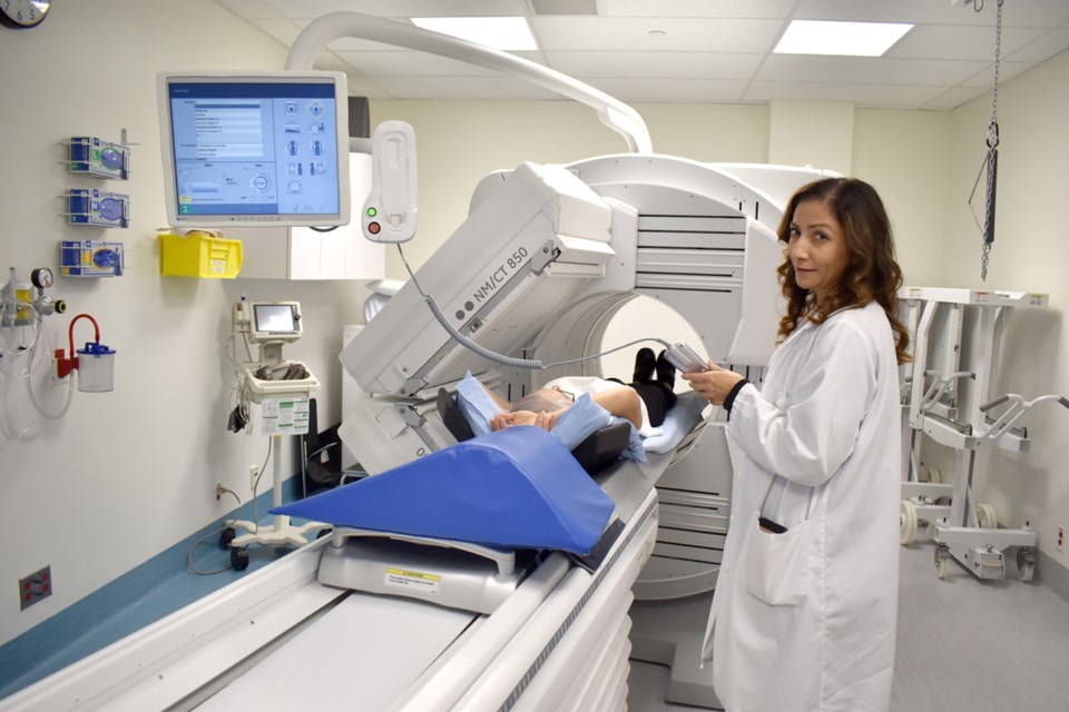
They are used for patients being diagnosed with the gamma camera – the latest diagnostic tool at the hospital and the first of its kind in Canada.
After its final installation this summer, the camera has been used 500 times for detecting medical problems in the lungs, heart, kidneys, gall bladder, bones, brain, thyroid gland and sometimes the stomach.
The camera cost $1.9 million – that covered renovating and expanding the room where it’s located, adding lead windows and upgrading the HVAC system. The Richmond Hospital Foundation gave $300,000 toward the new camera.
While the camera allows for very specialized diagnostics, Lesley Lee, supervisor of nuclear medicine at Richmond Hospital, said it also increases patients comfort by reducing time spent in the machine and allowing the patient to relax while the camera rotates around to take its pictures.
“It has higher image resolution than our previous camera, and it has built-in SPECT/CT capability, so it helps radiologists interpret results with greater confidence,” she said.
Unlike other specialized diagnostic tools, like MRIs and CT scans, the gamma camera can see the functionality of organs and pinpoint things like the nodes where breast cancer is located so they can be targeted for treatment.
Or, while some diagnostics show whether there are stones in a gall bladder, the gamma camera can show how well the gall bladder is performing.
Nuclear medicine is a subspecialty of diagnostic imaging, which involves the use of radioactive medications (radiopharmaceuticals – the isotopes) to diagnose and treat disease.
The radiopharmaceutical is administered by IV injection, swallowed, or inhaled, then tracked from outside the body using a gamma camera to detect the radiation it emits.
Depending on the type of procedure, two or three dimensional images of the internal body are created.


